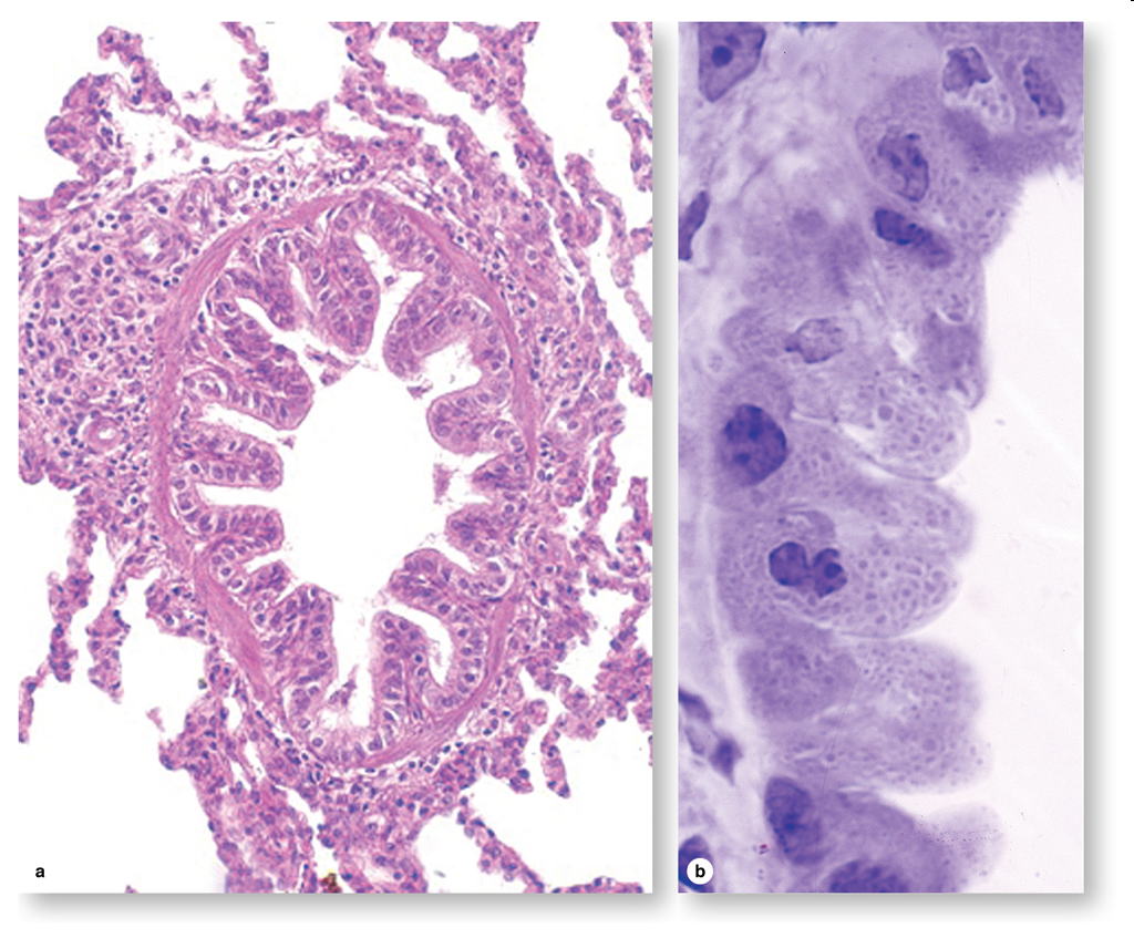

Figure 17-10.
Terminal bronchiole and Clara cells. The last parts of the air conducting
system before the sites of gas exchange appear are called the terminal
bronchioles, which generally have diameters of one to two mm. (a): Cross-section
shows that a terminal bronchiole has only one or two layers of smooth muscle
cells. The epithelium contains ciliated cuboidal cells and many low columnar
nonciliated cells. X300. PT. (b): The nonciliated Clara cells with bulging
domes of apical cytoplasm contain granules, as seen better in a plastic
section. Named for Dr. Max Clara, the histologist who first described them
in 1937, these cells have several important functions. They secrete components
of surfactant which reduces surface tension and helps prevent collapse
of the bronchioles. In addition, Clara cells produce enzymes that help
break down mucus locally. The P450 enzyme system of their smooth ER detoxifies
potentially harmful compounds in air. In other defensive functions, Clara
cells also produce the secretory component for the transfer of IgA into
the bronchiolar lumen; lysozyme and other enzymes active against bacteria
and viruses; and several cytokines that regulate local inflammatory responses.
Mitotically active cells are also present and include the stem cells for
the bronchiolar epithelium. X500. PT.