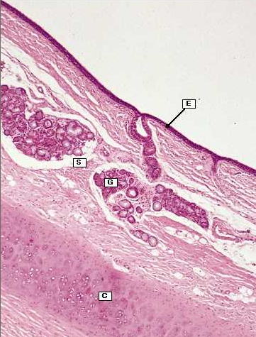
Fig. 10.8b Trachea.
Micrograph showing tracheal mucosa with ciliated respiratory epithelium (E) on its surface and seromucousglands (G) in the submucosa (S). The inner part of the tracheal cartilage ring (C) can be seen at the base of the micrograph.