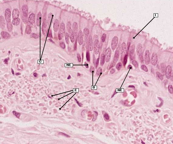
Fig. 10.10 Bronchial epithelium.
High-power micrograph of a thin acrylic resin section of the bronchial epithelium showing the various cell types present. Most of the cells are tall columnar ciliated cells (C) but
scattered between them are occasional intermediate cells (I). Goblet cells are not seen in this section, but basal (B) and neuroendocrine (NE) cells are present on the basement
membrane. Longitudinal elastic fibres (E) of the bronchial wall, here cut in transverse section, can also be seen.

